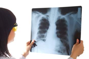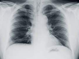
Chest X-Ray

Mesothelioma Diagnosis: Chest X-Ray Malignant mesothelioma is an infrequent cancer that primarily forms in the mesothelium, which is a network of tissues that shield several internal organs, most notably the lungs and heart. Mesothelioma cancer is often caused by prolonged exposure to asbestos filaments. Asbestos is a naturally occurring element that, when disrupted or contacted, propels carcinogenic fibers into the air. When inhaled, these filaments, stick to the mesothelium. Over time, these carcinogens eat away at the protective tissues and promote the formation of cancerous tumors. Because of the disease’s close link to asbestos exposure, the bulk of malignant mesothelioma patients will have a prolonged history with handling or working with asbestos-related products. Mesothelioma tumors, if malignant, will eventually metastasize to other organs. When this occurs, symptoms begin to arise, that although, minor in nature, and precipitate a quick death. Although there are multiple forms of mesothelioma cancer, the bulk of cases formulate in the lining of the lungs or the internal wall of the chest. Common symptoms attached to mesothelioma cancer include: shortness of breath (stems from a build-up of fluid between the lungs and abdomen), severe and unexpected weight loss and painful/persistent coughing. One of the principal characteristics attached to mesothelioma cancer--and the major reason why the cancer is so deadly--derives from its relatively harmless symptoms during the disease’s earliest stages. Mesothelioma cancer is practically impossible to detect even 15 years after the cancer has spread throughout the body. Therefore the problems associated with accurate diagnosis not only stem from the cellular structure of the disease (early-stage mesothelioma cells are difficult to detect), but also from the cancer’s lack of tangible symptoms. Mesothelioma patients will start noticing the aforementioned symptoms a ridiculous 20 years after the disease has stricken the body. Mesothelioma symptoms, on average, will become observable when the cancer reaches its 3rd or 4th stages. Similar to most cancers, malignant mesothelioma is categorized by stage. As malignant mesothelioma progresses into its 3rd and 4th stages it metastasizes and becomes highly aggressive and fatal. The insidious nature of mesothelioma cancer is paired with the disease’s inconspicuousness, to formulate one of the most lethal medical conditions on the planet. As a result, the majority of mesothelioma patients will typically fall victim to the cancer within 6 months to 1 year of diagnosis. In the bulk of cases, mesothelioma cancer will not be diagnosed until the mesothelioma tumors advance to the vital organs of the body. Stage III or Stage IV mesothelioma cancer is often deemed inoperable. If the cancer, by a small miracle, is detected before it spreads, curable mesothelioma treatments, such as surgery, may be applied to extract the cancerous tumors. If the disease is not detected in its early stages (which are more common) palliative treatment options, including surgeries, will be recommended to ease the pain and suffering associated with mesothelioma cancer. If you have worked or are working with asbestos fibers or asbestos-containing materials you should contact a medical professional to schedule a physical examination. This appointment should be made regardless of how you feel; remember, malignant mesothelioma cancer will not yield noticeable symptoms for the first 10-15 years of the disease’s life. During a physical examination, your doctor will first take a chest x-ray if he believes you are at risk of malignant mesothelioma cancer. The chest x-ray is a preliminary image; if the chest x-ray reveals any irregularities the doctor will request further tests, including CT scans, MRI’s and PET scans. Chest X-Rays to Help Diagnose Mesothelioma: A chest x-ray is a medal imaging technique that is primarily used to diagnose injuries to bones. Chest x-ray technology; however, may also be utilized to diagnose problems in a patient’s soft tissue, including the lungs. The chest x-ray therefore, can be used to diagnose lung cancer, pneumonia and mesothelioma cancer. A chest x-ray is used by medical professionals to help reveal any unusual thickening of the pleura and whether fluids have accumulated in the pleural space (pleural effusion). The chest x-ray will also reveal the presence of calcium deposits on the pleura. If the chest x-ray reveals any of these things, the doctor will invariably order further imaging testing. A chest x-ray to diagnose mesothelioma cancer will also show evidence of pleural plaques and lung scarring, which may indicate the presence of asbestosis—a distinct and separate disease from mesothelioma cancer that is asbestos related and usually nonmalignant. A chest x-ray for mesothelioma patients will inevitably show irregularities. However, a chest x-ray has limited usefulness because the image’s findings are nonspecific. The chest x-ray cannot delve deep into the tissues and reveal irregularities that are picked-up by more advanced imaging platforms. Chest x-rays are common diagnostic imaging tools for mesothelioma cancers. A chest x-ray for diagnosing mesothelioma cancer is effective in pinpointing the location of a mesothelioma tumor. This ability enables the medical professional to observe whether or not the mesothelioma tumor has metastasized and spread to other areas outside of the pleural cavity. Chest x-rays, along with blood tests and CT scans are the preliminary diagnostic tools to detect the presence of mesothelioma cancer. However, the downside of these medical procedures is that are incapable of bolstering the diagnosis of mesothelioma cancer during the disease’s earliest stages of development. Chest X-Ray as a Treatment Option for Mesothelioma: There are three primary standards of care for mesothelioma patients. Chemotherapy, for one, can be utilized with a combination of medications to kill cancerous tumor cells. Surgery is another option to treat mesothelioma; these surgeries involve extracting the tumor and part or all of the mesothelium as well. The last common method used to treat mesothelioma is radiation. Radiation therapy is a severe dose of x-rays, which are directed to the cancerous cells. Radiation therapy (also referred to as x-ray therapy) is used to target and destroy individual tumor cells. As the cancerous cells die, the main tumor shrinks. A high dose of x-rays will be administered as a mesothelioma treatment option in a variety of ways. For instance, radiation therapy can be applied with a machine that is directed to a cancerous tumor externally. Moreover, radioactive materials can be administered in contained plastic shells and surgically implanted on the tumor. A medical professional may apply one or all three of these methods to treat mesothelioma cancer. A combined mesothelioma treatment package; however, will only be administered to mesothelioma patients who are deemed strong enough to sustain the cruel side effects imposed by these aggressive treatment options. Side Effects of Mesothelioma Chest X-Rays: There are several dangers associated with high level or long term exposure to x-rays. When a human body is exposed to x-rays, radiation occurs on a cellular level. In general, the radiation will damage cells, causing them to die or sustain permanent damages to their genetic makeup. This may eventually perpetuate the formation of cancerous cells. It may seem odd to treat cancer with a technology that may cause another, but x-rays and radiation therapy has proven effective in shrinking tumors. Presently, these techniques yield few side effects; doctors can now pinpoint and fine-tune the levels of radiation a patient will receive. Moreover, innovations enable the medical professional to better monitor the patient’s health, while using other therapies to reduce the side effects associated with chest x-rays and radiation therapy. Radiation therapy--consisting of high powered doses of x-rays--to treat mesothelioma cancer has several advantages over chemotherapy and other mesothelioma treatment options. That being said, chest x-rays and radiation may spark several disadvantages. These disadvantages widely stem from the fact that radiation therapy targets only areas where the tumor is located. Although this characteristic is effective in isolating cancer, it perpetuates the formation of several skin problems, including soreness, tightness and severe irritations. Moreover, radiation in the chest may make it difficult to swallow, which promotes severe coughs and shortness of breath just a few months after receiving treatment. The majority of side effects associated with radiation in the chest are temporary. If the patient experiences severe side effects, the medical doctor professional prescribe alternative medications, such as antibiotics or steroids to reduce the severity of said effects.


